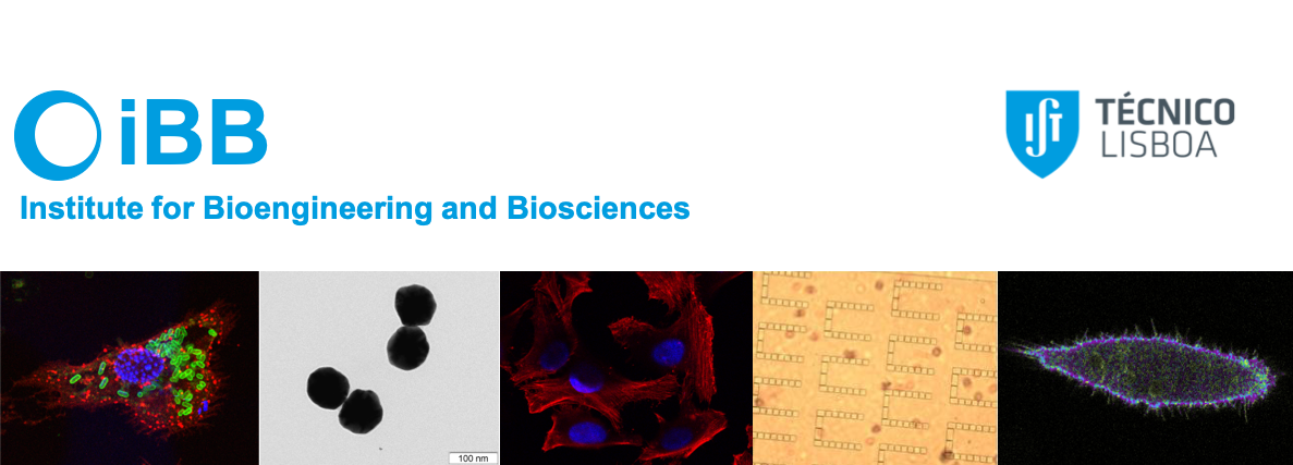
PPBI-Portuguese Platform of BioImaging
- Förster Resonance Energy Transfer (FRET) Microscopy;
- Two-photon microscopy;
- Fluorescence recovery after photobleaching (FRAP);
- Fluorescence lifetime imaging (FLIM, time-domain) and FLIM-FRET;
- Fluorescence correlation spectroscopy (FCS);
- Fluorescence cross-correlation spectroscopy (FCCS);
- Fluorescence anisotropy imaging microscopy (FAIM);
- Single-molecule FRET;
The team attained international recognition in molecular and cell biophysics. Remarkably, we implemented for the first time FAIM and single-molecule FRET in Portugal. The unit also provides scientific support in project development, technical training, and guidance in image acquisition/ analysis for iBB researchers and also academic visitors or companies. The facility is a node of the PPBI-Portuguese Platform of BioImaging, part of the National Roadmap of Research Infrastructures (funded by FCT).
 |
Resources Microscope #1: Leica SP5 Multiphoton/Confocal Fluorescence Microscope for distinct applications: • colocalization of fluorescent molecules and 3D sectioning/reconstruction; • spectral imaging; • two-photon microscopy; • functional imaging (FRET, FRAP, FLIM, FAIM, FCS and FCCS); It is equipped with: • CW lasers: Ar ion laser (458, 465, 488, 496 and 514 nm) and HeNe (633 nm); • Pulsed laser: Ti:Saphire (710-990 nm, 100 fs, 80 MHz); • Objectives: HCX PL APO 10x/0.4 Dry CS; HC PL FLUOTAR 50x/0.8 Dry; and HCX PL APO 63x/1.20 W (collar correction); • Microinjection: combination of InjectMan® 4 and Femtoject® 4i from Eppendorf; • Autocorrelator and two APDs for single-molecule FCS and FCCS; • TCSPC module coupled to a DCC-100 PMT or two hybrid detectors; Microscope #2: Olympus IXplore IX83 Widefield Microscope equipped with: • Digital Hamamatsu CMOS camera (C11440-36U); • Imaging Workstation (HP Z2 SFF G4); • cellSens dimension software; • Filter cubes: (1) Exciter filter: 340-390 nm, beam splitter: 410 nm, barrier filter: 420 nm; (2) Exciter filter: 470-495 nm, beam splitter: 505 nm, barrier filter: 510 nm; (3) Exciter filter: 530-550 nm, beam splitter: 570 nm, barrier filter: 575-625 nm; • Objectives: UPLXAPO10X and UPLXAPO60XO; Microscope #3: Home-built confocal microscope for single-molecule FRET: It is based on an inverted microscope (Olympus IX-73) containing a high numerical aperture objective (60x/1.2NA water immersion objective) and 2 APDs detectors (SPCM-AQR-14 from Excelitas). It integrates a Sapphire LP 488 diode laser (Coherent), 50 or 100-μm diameter optical fibers to achieve confocal aperture; and a Correlator Flex03LQ-12. Image analysis softwares: MATLAB, ImageJ and Imaris |
|
 |
Team Ana M. Melo - Senior Researcher and Facility Manager Ana Coutinho - Senior Researcher Fábio Fernandes - Senior Researcher Manuel Prieto - Senior Researcher Mario Berberan-Santos - Senior Researcher Sandra Pinto - Senior Researcher |
|
 |
Services Please contact Ana M. Melo |
|